Mammography in Lausanne
Search and Compare the Best Clinics and Doctors at the Lowest Prices for Mammography in Lausanne

Find the best clinics for Mammography in Lausanne
No pricing info available
Malaysia offers the best prices Worldwide
Price: $ 11
From 10 verified reviews
interprète ? Couyère, 24 January 2020
this morning in radiology, all very nice and very efficient I recommend 👍
From 21 verified reviews
Doriana RIVA, 10 September 2020
A huge thank you to the healthcare team who took care of me urgently on April 22, 2020. The welcome at the front desk, the anesthesia team, the assistants, the nurses in the operating room and the recovery room were so sweet and very attentive.I will never forget that time spent with you. Good luck to everyone, e.D. Riva
- Home
- Switzerland
- Lausanne
WHY US?
At Medijump, we're making medical easy. You can search, compare, discuss, and book your medical all in one place. We open the door to the best medical providers worldwide, saving you time and energy along the way, and it's all for FREE, no hidden fees, and no price markups guaranteed. So what are you waiting for?

Free

Best Price

Widest Selection

Risk-Free
What you need to know about Mammography in Lausanne

Mammography is an X-ray for the breast. It is used to detect and diagnose breast cancer and other breast diseases. The images that mammography produces is called mammograms. The images can show tiny tumors that cannot be felt, as well as other abnormalities in the breast.
Mammography can be used for screening and diagnostic purposes:
-
Screening mammography looks for breast cancer, benign tumors, cysts, or other breast diseases in women with no symptoms. The goal of the procedure is to detect the disease early, when it may be more treatable.
Diagnostic mammography is usually done for women who have symptoms of breast cancer or who have a high risk of developing the disease. It may be recommended if you feel a lump in the breast, you experience other unfamiliar symptoms, or a screening mammogram shows a suspicious area.
What does a Mammography Procedure Involve?
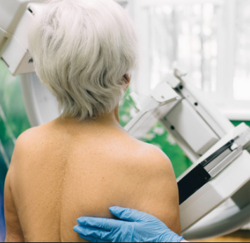
During mammography, you will either stand or sit in front of the mammography machine. Your breast is placed on a flat support plate. Then, a compressor will push the breast down to flatten it. Flattening your breast is done to spread out the tissue, which can provide clearer pictures and make it easier to find smaller abnormalities.
Once your breast is flattened, your doctor or technician will take an X-ray image. You may have to hold your breath to reduce the possibility of a blurred image while the X-ray image is taken. Your doctor may also ask you to change positions for each picture. You may feel a little discomfort or pressure, but it is usually brief.
When X-ray images are taken, a small burst of x-rays passes through your breast to a detector that is located on the opposite side. This detector is usually a photographic film plate that can capture the X-ray image on film. However, today, many breast imaging centers have moved from using film plates to digital mammography. Here are the newest advances in mammography:
-
Digital mammography – digital mammography records the images on a computer instead of on film. A solid-state detector is used to transform the X-ray into a digital image that saves onto a computer. The computer can help your doctor see images that might not have been very visible on a regular mammogram because the image contrast is sharper. Digital mimeographs are also easier to store and share with other medical professionals.
-
3D Breast Imaging – this is called breast tomosynthesis. For this test, the breast is positioned and flattened just like a traditional mammogram. However, a tomosynthesis uses an X-ray tube that moves in an arc and takes pictures of your breast from many angles. Several studies have shown that this test, results in improved breast cancer detection rates, but it is not yet widely available.
Once the test is complete and all the necessary images have been taken, a radiologist will carefully examine the mammogram.
How Long Should I Stay in Lausanne for a Mammography Procedure?
You can leave the hospital immediately after your mammography is complete. However, since it may take at least a week until the result is complete, it is advisable that you stay in Lausanne for at least 7 days. Once the result is ready, you need to attend a follow-up appointment to discuss the result with your doctor. In some cases, it may take several weeks until the results are ready. If this is the case, talk to your doctor/medical travel team about the exact length of stay, and the results can be mailed/sent to you.
What's the Recovery Time for Mammography Procedures in Lausanne?
You can resume your normal activities, including work, right after your mammogram. Some women experience minor bruising, but most women do not feel any lingering pain at all once the pain is over.
What sort of Aftercare is Required for Mammography Procedures in Lausanne?
If you experience soreness, minor bruising, or discomfort, you should wear a padded sports bra as it can help you feel more comfortable then wearing a bra with underwire.
Although visible bruising on your breast or soreness a full day after your mammogram takes place is not cause for alarm, you should let your doctor know.
What's the Success Rate of Mammography Procedures in Lausanne?
Mammograms are considered as one of the best breast cancer screening tests currently available. However, they have their limits as they are not 100% accurate in showing if a woman has breast cancer. There is always a possibility of:
-
A false-positive – the mammogram looks abnormal even though there is actually no cancer cells present in the breast.
-
A false-negative – the mammogram looks normal even though there is breast cancer.
Besides accuracy, the procedure itself is very safe. It should not cause alarming or long-term side effects as the amount of radiation used is minimal.
Are there Alternatives to Mammography Procedures in Lausanne?
If you cannot or do not want to undergo mammography, you can consider the alternatives. These include ultrasound, MRI, and molecular breast imaging (MBI). However, mammograms are still considered as the best screening test for breast cancer since none of the alternatives has been proven to be as good.
What Should You Expect Before and After the Procedure
After mammography, your doctor can check the condition of your breast, whether or not you experience any symptoms before. If the results show any abnormalities (positive), your doctor may suggest you undergo a breast biopsy and they will be able to create a treatment plan.
Whilst the information presented here has been accurately sourced and verified by a medical professional for its accuracy, it is still advised to consult with your doctor before pursuing a medical treatment at one of the listed medical providers
No Time?
Tell us what you're looking for and we'll reachout to the top clinics all at once
Enquire Now

Popular Procedures in Lausanne
Prices Start From $42

Prices Start From $4

Prices Start From $23

Prices Start From $11

Recommended Medical Centers in Lausanne for Mammography

- Interpreter services
- Translation service
- Religious facilities
- Medical records transfer
- Medical travel insurance
- Health insurance coordination
- TV in the room
- Safe in the room
- Phone in the room
- Private rooms for patients available

- Interpreter services
- Translation service
- Religious facilities
- Medical records transfer
- Medical travel insurance
- Health insurance coordination
- TV in the room
- Safe in the room
- Phone in the room
- Private rooms for patients available

- Interpreter services
- Translation service
- Religious facilities
- Medical records transfer
- Medical travel insurance
- Health insurance coordination
- TV in the room
- Safe in the room
- Phone in the room
- Private rooms for patients available

- Interpreter services
- Translation service
- Religious facilities
- Medical records transfer
- Medical travel insurance
- Health insurance coordination
- TV in the room
- Safe in the room
- Phone in the room
- Private rooms for patients available

- Interpreter services
- Translation service
- Religious facilities
- Medical records transfer
- Medical travel insurance
- Health insurance coordination
- TV in the room
- Safe in the room
- Phone in the room
- Private rooms for patients available

- Interpreter services
- Translation service
- Religious facilities
- Medical records transfer
- Medical travel insurance
- Health insurance coordination
- TV in the room
- Safe in the room
- Phone in the room
- Private rooms for patients available

- Interpreter services
- Translation service
- Religious facilities
- Medical records transfer
- Medical travel insurance
- Health insurance coordination
- TV in the room
- Safe in the room
- Phone in the room
- Private rooms for patients available

- Interpreter services
- Translation service
- Religious facilities
- Medical records transfer
- Medical travel insurance
- Health insurance coordination
- TV in the room
- Safe in the room
- Phone in the room
- Private rooms for patients available

- Interpreter services
- Translation service
- Religious facilities
- Medical records transfer
- Medical travel insurance
- Health insurance coordination
- TV in the room
- Safe in the room
- Phone in the room
- Private rooms for patients available

- Interpreter services
- Translation service
- Religious facilities
- Medical records transfer
- Medical travel insurance
- Health insurance coordination
- TV in the room
- Safe in the room
- Phone in the room
- Private rooms for patients available
Mammography in and around Lausanne
Popular Searches
- Plastic Surgery in Thailand
- Dental Implants in Thailand
- Hair Transplant in Thailand
- Breast Augmentation Thailand
- Gastric Sleeve in Thailand
- Gender Reassignment Surgery in Thailand
- Laser Hair Removal in Bangkok
- Botox in Bangkok
- Dermatology in Bangkok
- Breast Augmentation in Bangkok
- Coolsculpting in Bangkok
- Veneers in Turkey
- Hair Transplant in Turkey
- Rhinoplasty in Turkey
- Stem Cell Therapy in Mexico
- Rhinoplasty in Mexico
- Liposuction in Mexico
- Coolsculpting in Tijuana
- Rhinoplasty in Korea
- Scar Removal in Korea
- Gastric Sleeve in Turkey
- Bone Marrow Transplant in India
- Invisalign in Malaysia
- Plastic Surgery in the Dominican Republic
- Tummy Tuck in the Dominican Republic
- Plastic and Cosmetic Surgery in Poland
- Rhinoplasty in Poland
- Hair Implant in Poland
- Dental Implants in Poland
- IVF in Turkey

